Sunday, December 11, 2011

Rhinoplasty: risks and pitfalls

Complications of Rhinoplasty
S.Valentine Fernandez

The operation of rhinoplasty demands a thorough understanding of an art and science.Some complications of rhinoplasty relate to anesthesia and do not fall within the bounds of this discussion.

Complications of rhinoplasty may be divided into 4 basic categories as follows: 
  • Intraoperative
  • Immediate post operative (in the recovery ward)
  • Early postoperative
  • Late postoperative
While recognizing obvious medical and surgical complications should present no difficulty, finer aesthetic complications are harder to define and are based on value judgments related to the aesthetic sense of the surgeon and the body-image demands of the patient Aesthetic sense is difficult to define, and it is much harder to agree on results. Aesthetics depend on variables, including the current fashion taste, the media, the public relations industry, and cultural and ethnic differences.

A practical approach to aesthetics in the nose begins with an accurate assessment. Frontal views define x-axis (width) and y-axis (height) deformities, lateral views define z-axis (depth/projection) and y-axis deformities, and basal views define x-axis and z-axis deformities. Based on these views, a 3-dimensional concept of the nose is made available for manipulation. The goal of rhinoplasty is to improve the existing harmony without causing functional impairment.

According to the literature, the complication rate for nasal surgery varies from 4-18.8%. In individual hands, this rate generally falls as surgical experience accumulates. Skin and associated soft tissue complications occur in up to 10% of cases. According to estimates, severe systemic or life-threatening complications occur in 1.7-5% of rhinoplasty cases. Intracranial complications are rare.

The clinical manifestation of rhinoplasty complications may broadly be classified as follows:
  • Functional
  • Infectious
  • Aesthetic
  • Psychological
  • Specific to complication       
Intraoperative complication:
1. Excessive bleeding  2. Tears of mucoperichondrial flaps  3. Buttonholing of skin  4. Cautery burns

5. Collapse of bony pyramid:     
  • Collapse of the bony pyramid may occur during removal of a bony hump with an osteotome, particularly when the patient has had previous nasal trauma or if the vomer or ethmoid have been weakened as a result of previous surgery. Rasping may be advisable in these circumstances.
  • Rectification requires careful approximation of the segments and provision of adequate internal and external splint support during healing. 
6.  Disarticulation of upper lateral cartilage:  This complication may occur during rasping. Bilateral disarticulation produces an inverted-V deformity, and unilateral disarticulation produces asymmetry in the middle third of the nose. Spreader grafts may improve airway symptoms and aesthetics.

7. Osteotomy complications 
  •  "Rocker" deformity: This deformity results when the medial osteotomy creates a cephalic fracture higher in the thicker part of the frontonasal junction. Attempted narrowing after the osteotomies results in lateralization of the superior segment of the fractured bones, based on a fulcrum at or about the radix. Repositioning the cephalic fracture lower on the nasal bone rectifies this deformity.
  • "Open roof" deformity: When the lateral segments fail to align with the septal dorsum following osteotomies, a gap, which may be visually and palpably obvious, results. If neglected, the intranasal mucous membrane adheres to the overlying soft tissue and may create a depression at the site. If alignment failure is unilateral, the nose appears asymmetric. Correction involves assuring centralization of the septum and complete medial mobilization of the lateral segments following osteotomy. Usual causes of open roof deformity include the following:  
    • Greenstick cephalic fracture during osteotomies (will return to its preoperative position)
    • Failure to adequately mobilize the fractured segments medially
    • Excessive nasal packing
    • Uncorrected deviated perpendicular plate of the ethmoid (may prevent medialization of the lateral segments)
  • "Step" deformity: This deformity may result if a single lateral osteotomy is performed too far medial to the nasofacial groove, with a visible ridge on the side of the nose. Correction involves repeating the osteotomy at the correct level. 
8. Perinasal trauma: During osteotomy, particularly in noses that have previously been traumatized, there is a tendency to recreate preexisting fractures. The proximity of the orbit and cranium renders these structures most susceptible to such occurrences. Malfunction can result directly or can manifest subsequent to infection.

Immediate post operative complications: 
1. Airway obstruction 2 Anaphylaxis: This is a distinct possibility when intraoperative antibiotic medication is used. Anaphylactic shock subsequent to bacitracin nasal packing has been reported, and latex anaphylaxis is well document 3. Visual impairment

Early post operative complications:                                                              
1. Hemorrhage: Reported prevalence of hemorrhage varies from 2-4%.   2. Septal hematoma:

3. Infection:  Wound infection, Septicemia, Toxic shock syndrome, subacute bacterial endocarditis, intracranial infections, and acute &/or chronic sinusitis.

4. Dehiscence of incisions: The transcolumellar incision must be attended to immediately, or a scar will form, contributing to a poor result.

5.Persistent edema: Persistent edema and numbness over the nasal tip region may occur following external rhinoplasty and may last several months. This is not a problem if the patient has been forewarned.

6. Skin necrosis: Excessive undermining, injudicious cautery use, and overzealous skin thinning may lead to skin necrosis. This may cause an impaired blood supply and infection. Similarly, a tight dressing may cause vascular impediment and skin necrosis. Debridement and secondary healing is encouraged if necrosis occurs. Later, local steroid injection, dermabrasion, laser modification, and/or flap reconstruction may be necessary to aid scar aesthetics. Skin necrosis may also result from dorsal augmentation, in which case removal of the augmenting material may be necessary.

7. Sequestra formation, 8. Cardiovascular insufficiency 9. Cerebrospinal fluid rhinorrhea 10. Contact dermatitis 11. Nasal blockage 12. Numbness and pain 13. Olfactory disturbance:  Patients with past facial trauma may be predisposed to injury of the olfactory apparatus during osteotomy. 14. Carotid-cavernous fistula 15. Reassurance demand:  A small number of patients need the surgeon to repeatedly express that the nasal blockage will disappear, the smell and taste sensation will return, the teeth anesthesia will subside, and the tip projection and swelling will decrease in time.Early psychological complications: Transient episodes of anxiety or depression are not uncommon and may last up to 6 weeks after the operation

Late postoperative complications

1. Scar hypertrophy: This may detract from a good result following an external rhinoplasty. Skin loss from infection and necrosis is a disaster. Aim initial attempts at reducing the size of the scar with intralesional steroids. Further treatment could include dermabrasion, lasers, and/or surgical scar revision.
2. Polly beak nasal deformity-This deformity is characterized by absence of the supratip dip (ptosis tip)  may present in degree.

3. Synechiae formation  4. Septal perforation: The prevalence of this complication has been described as 3-24.5%.

5. Nasal valve collapse: Aggressive cephalic trim of the lower lateral cartilages may provoke this complication. Collapse may cause airway distress and is a source of patient discontent. Cartilage spreader grafts may prove useful for internal valve collapse. Alar batten grafts improve external valve collapse.

6. Nasal stenosis  This is a disaster when it occurs, and it may be related to circumscribed incisions with excessive lining removal. Stenosis causes airway obstruction and is a source of persistent discomfort. Reconstructive surgery may provide some relief.

7. Bossa formation
  • Bossae are protuberances that may arise in the region of the nasal tip. Their postoperative incidence is quoted at 2%.
  • While bilateral symmetric occurrences may assume a pleasing appearance, bilateral asymmetric and unilateral bossae demand surgical attention. During operation, ensuring that the remnant lower lateral cartilages are of equal dimensions on both sides is important.
  • Bossae usually occur in noses with preoperative asymmetry, use of destructive rhinoplasty techniques, thin skin, and excessive postoperative scarring. The triad of thin skin, strong cartilages, and bifidity also indicate the patient who is prone to the development of bossae. Tip graft movement may also contribute to bossa formation. 
8. Recurrent meningitis, 9. Oleogranuloma  10. Dorsal cyst:

11. Aesthetic surgical misjudgments
  • Undercorrection or overcorrection of a preexisting deformity leads to either persistence of the deformity or to introduction of a new one. A new deformity may introduce a functional deficit. Some of these deformities are illusory, and correction only follows after an accurate diagnosis is made. Ideally, revision rhinoplasty should not be performed until at least 12 months after the initial operation.
  • These deformities may occur singly or in combination and may relate as an x-axis (width), y-axis (height), or z-axis (depth) deformity/deformities in the various segments.

Upper third deformities 
Deep nasofrontal angle, Shallow nasofrontal angle, Upper third widening, Upper third convexity, Upper third overreduction:,  & Upper third asymmetry.

Middle third deformities 
Middle third widening:, Middle third convexity, Middle third saddling,  & Middle third asymmetry

Lower third deformities
Lower third widening or flaring,  Tip widening or boxy tip, Tip narrowing or pinched tip, Tip asymmetry,   Tip projection deformities, Wide columella, Hanging columella or "columella show", "Hanging" or "veiled" alae , Alar notching, Nostril asymmetry, Retracted nasolabial angle, Protracted nasolabial angle,   &

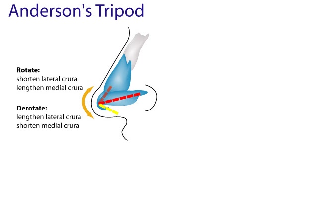
Alar collapse: Excessive removal of the lateral crura dampens support afforded by these structures, causing alar collapse and airway discordance. Reconstructive techniques with alar battens may be necessary in difficult cases.

Other

  • Graft/implant migration: Migration may comprise resorption, displacement, or extrusion; it may be provoked by trauma and infection. Allografts have a higher extrusion and infection rate than autografts. An infected implant must be removed if antibiotics do not help. Revise displaced grafts causing aesthetic inconvenience with appropriate stabilization.
  • The disproportionate nose: This nose does not fit the face and is not a credit to the surgeon's artistic skill. Reconstructive rhinoplasty techniques follow the patient's expression of discontent with original results.
  • Underlying maxillofacial deformity: A superb rhinoplasty result may be ruined by the unmasking of a previously unnoticed or undiagnosed maxillofacial deformity. The maxillae and mandible with labial and dental components must be considered preoperatively and the patient must be forewarned. Attendance by a cosmetic dentist and inclusion of chin surgery may be necessary.
A revision rhinoplasty may be required in 5-15% of patients. Any attempt at correction must always be accompanied by a rider promising only improvement and warning that a further "touch-up" operation may be necessary.

Persistent psychological complications: Several contradictory studies are available.
Thoracic and thoracolumbar instability, Gustatory rhinorrhea, Human adjuvant disease:, Lacrimal fistula, Enophthalmos and silent sinus syndrome. 

For more detailed analysis see:
http://emedicine.medscape.com/article/843439-overview#showall

Some other complications not mentioned above: Neuropathic pain (temporary or permanent),  Empty Nose Syndrome, Graft warping and resorption and complications from injectable material in the nose. For more info on this subjects check out  my other blog posts. (See labels on right side column of my blog and click on any of the labels.)

See also:
http://www.youtube.com/watch?v=sj4RrAlAwXo
http://yourguidetoplasticsurgery.com/rhinoplasty-risks
http://www.emptynosesyndrome.org/what_is_ens.php
http://www.therhinoplastycenter.com/ComplicationsofRhinoplasty/complications1.html

    Saturday, December 10, 2011

    Refining the nasal tip with Newer VDD techniques.

    Vertical Dome Divison Rhinoplasty: 
     Author: John Hilinski, CoAuthors: Anil R. Shah & Kris S Moe

    Nasal tip surgery is among the most complex and difficult tasks in rhinoplasty surgery. Vertical dome division (VDD) is one of a variety of techniques that may be used in refining nasal tip appearance. Collectively, vertical dome division (VDD) refers to one of many methods of vertically dividing the lower alar cartilage at or near the dome to modify nasal tip aesthetics. The technique was originally recommended as an alternative in altering tip projection and appearance while minimizing use of implants and the degree of postoperative tip ptosis. Vertical dome division (VDD) targets various nasal deformities, including overprojection or underprojection, suboptimal rotation, disproportionate lobule ratios, and broad or asymmetric tip. The surgeon must strive to achieve an aesthetically pleasing nasal tip that is in balance with the remainder of the nose without compromising nasal airway function.

    The typical patient presenting for vertical dome division (VDD) usually has a poorly defined or malpositioned tip with a combination of abnormal projection/rotation, broad or amorphous lobule, asymmetric tip defining points, and/or boxy, trapezoidal base.

    Adherents to this principle argue that horizontal excisional techniques rely too heavily on unpredictable and uncontrollable postoperative scarring to produce desired tip results. Proponents of vertical dome division (VDD) believe that vertical incisional and excisional techniques, on the other hand, offer a more definitive and reliable means to achieve desired tip changes.

    In principle, the technique of vertical dome division (VDD) separates the medial and lateral crura into 2 independent units. By transecting the dome, the inherent spring within the arch is released and allows realignment of the newly divided medial and lateral segments to reconstruct the nasal tip.

    Vertical dome division (VDD) is typically reserved for more complicated cases that require greater changes to effect tip refinement than could be achieved using other techniques. Nearly all variations of vertical dome division (VDD) used today involve some modification of the original Goldman technique

    The lobule is defined as the portion of the nasal tip complex that is situated anterior to the nostrils; it extends from the tip defining point to the junction with the columella, as observed on base view. The alar cartilage (lower lateral cartilage) is C-shaped and can be divided into the medial, middle, and lateral crus. The middle (intermediate) crus comprises the domal segment and largely influences the shape of the lobule and, therefore, the form and definition of the nasal tip. The dome is considered the highest arching segment within the nasal vestibule.

    The lobule size can be assessed in comparison to the columellar length. If the base view demonstrates a columellar-to-lobule ratio of approximately 2:1, the structural support and configuration of the nasal tip is considered adequate. A long nasal length reflects an acute nasolabial angle; a short length reflects an obtuse nasolabial angle.

    The anatomy of the nasal tip is often described using the tripod concept to facilitate understanding of the key structural components and to provide a simple explanation of tip dynamics. According to this analogy, the cartilaginous framework of the lower third of the nose is compared to a tripod that is attached to the facial frontal plane. The 2 individual lateral crura represent 2 legs of the tripod, and the conjoined medial crura and caudal septal attachments correspond to the third leg.

    By lengthening or shortening any or all legs of the tripod, the changes that will be effected in tip projection and rotation can be predicted. For instance, techniques that augment or lengthen the medial crural segment enhance projection. Shortening the medial crura or disrupting their septal attachments without reduction of lateral crural length decreases projection and rotation of the nasal tip. Shortening the lateral crura and maintaining or lengthening the medial crural segment would be expected to increase rotation.

    Contraindications

    Vertical dome division (VDD) is predominantly contraindicated in patients with relatively thin skin. These patients are particularly prone to developing visible cartilage edges along the nasal tip region. This results from contraction of the thin overlying skin and soft tissue envelope around the new and more prominent medial cartilaginous strut. A thick overlying skin and soft tissue envelope is better able to cushion the appearance of prominent cartilaginous structures, such as those in vertical dome division (VDD).

    Avoid classic vertical dome division (VDD) in patients who show evidence of already weakened lateral nasal walls. Dividing the domal region without reapproximation of the cartilage segments disrupts the integrity and continuity of the lower lateral cartilage. The lateral nasal wall is more susceptible to structural collapse than the newly reinforced medial footplates. As a result, lateral wall weakening and collapse are further potentiated.

    The original technique is highly focused on manipulation and repositioning of only the medial crura, with no attempt made to reconstruct the remaining lateral crural segment and alar rim. Postoperatively, the medial crura and columella are sufficiently stable to resist loss of projection. Some loss of lateral support, which could result in lateral wall collapse and alar retraction, may occur. Vertical dome division (VDD) is also associated with bossae formation. This is most frequently seen in patients with thin skin and firm cartilages. Disruption of the underlying vestibular mucosa and skin, such as in the Goldman technique, also predisposes the patient to possible stenosis.

    With use of newer modified techniques (Hockey stick excision, Lipsett, Simons, Adamson, and others) and caution, vertical dome division (VDD) can be used successfully in nasal tip refinement with limited postoperative complications and reliable long-term results. 

    Most practitioners using vertical dome division (VDD) today perform some modification of the original Goldman tip procedure.

    Adamson reported that approximately 5% of patients required revision surgery for postoperative tip abnormalities and irregularities attributed to use of vertical dome division (VDD).[10] These abnormalities were primarily nasal bossae and lobule asymmetries. Abnormalities were nearly 3 times as likely to occur in revision cases as in primary rhinoplasty; incidence was lower with use of the incision and overlap method.

     Vertical dome division (VDD) is likely to remain controversial in the future. As knowledge of nasal tip surgery evolves, so too will further modifications of our existing tip techniques to attain more predictable outcomes.
     http://emedicine.medscape.com/article/841313-overview

    The Endonasal Approach to Rhinoplasty
    Robert L. Simons and Lisa D. Grunebaum Ch.68 Rhinology and Facial Plastic Surgery 

    Today's trends in nasal tip surgeries are for improved visualization with preservation of the cartilage as well as better medial stabilization and support. These tenets are inherent in vertical dome division techniques. It is important to remember that VDD is an incisional technique that allows for repositioning of the nasal tissue and should not be combined with excisional techniques. One should always leave behind more than one takes. Preservation of at least 6-8mm of lateral crus will help prevent alar collapse and help stabilize the nasal base as well as allow for a strong natural-appearing tip.VDD allows for narrowing, rotation, and change in tip projection by repositioning rather than excision of any sizable amount of cartilage.


    The Effectiveness of modified VDD Technique in reducing nasal tip projection in rhinoplasty
    B.Gandomi, M.H. Arzaghi, M. Rafatbakhsh

    The employed technique is a new modification of previous techniques. 3-10 The technique employs an open approach in which a strong columellar strut inserted and a portion of crura is removed near the dome (the cornerstone of our new technique of tip surgery). Depending on the deformity, this segment may involve intermediate, middle or lateral crura with or without removal of vestibular skin considering its thickness: thin skins are not resected, but thick skins are usually removed.

    Our technique involves the overlapping of the incised edges of the medial and lateral segments, and suture approximation to restore the integrity of the alar cartilage. The technique allows a more stable configuration for the maintenance of nasal tip support. The overlapping and reapproximation of the medial and lateral units ensure the long-term stability of the newly reconstructed nasal tip complex, and reduce the tendency towards postoperative cartilaginous abnormalities that may accompany scar fibrosis and contracture. 


    Wednesday, June 22, 2011

    Nasal tip complications and optional operative fixes

    Complications in Rhinoplasty-Daniel G. BeckerCh.49 facial plastic and reconstructive surgery by Ira D. Papel

    In the nasal tip;  

    Over-reduction may violate critical tip support mechanisms (Table 49.1) which can lead to complications including tip ptosis and inadequate tip projection. Alternatively, overresection of the caudal septum can result in overrotation of the nasal tip with excessive shortening of the nose. Overresection may also contribute to other complication such as bossae, alar retraction, and alar collapse (external nasal valve collapse).

    Under-reduction may be simply due to overcaution but is commonly due to a failure to correctly assess preoperatively the anatomical situation. For example, failure to recognize an overprojected nose, or to diagnose the steps required based on the patient's anatomy to adequately address this, can lead to a persistent overprojected state. Failure to adequately resect cartilaginous dorsum may result in a pollybeak deformity.

    Asymmetries of the nasal tip may be due to unequal reduction of the lower lateral cartilages or to asymmetric application of dome-binding sutures. It may also be caused by unequal scarring that can occur during the natural healing process and may not be evident for months or even years after surgery. Also, assymmetry is often present preoperatively and should be recognized and pointed out to the patient prior to surgery.

    Table 49.1 Tip-Support Mechanisms

    Major tip-support mechanisms
    1. Size, shape, and strength of lower lateral cartilages
    2. Medial crural attachment to caudal septum
    3. Attachment of caudal border of upper lateral cartilages to cephalic border of lower lateral cartilages
    [the nasal septum is also considered a major support mechanism of the nose]

    Minor tip-support mechanisms
    1.Ligamentous sling spanning the domes of the lower lateral cartilages (i.e. interdomal ligament)
    2.Cartilaginous dorsal septum
    3.Sesamoid complex of lower lateral cartilages
    4.Attachment of lower lateral cartilages to overlying skin-soft tissue envelope
    5. Nasal spine
    6. Membranous septum

    Specific Complications:

    Ptotic tip-loss of tip support may lead to a droopy tip (tip ptosis with an overly acute nasolabial angle (angle defined by columellar point to subnasale line intercepting with subnasale to labrale superious line) is 90 to 120 degrees.Within this range, a more obtuse angle is more favorable in females, a more acute angle in males. Loss of tip support can lead to a ptotic, underprojected drooping nose. Treatment of complications relating to a ptotic nose rely on restoration of tip support and tip projection.

    Overrotated Tip-Conversely, one may face a patient with a nose that has been overrotated, with an overly obtuse angle. Overresection of the caudal septum is a common cause of overrotation of the tip. Overrotation of the nose creates an unsightly, overshortened appearance. Treatment rely on maneuvers that lengthen and counterrotate the nose.

    Bossae- a bossae is a knuckling of the lower lateral cartilage at the nasal tip due to contractural healing forces acting on weakened cartilages. Patients with thin skin, strong cartilages, and nasal tip bifidity are especially at risk. Excessive resection of lateral crus and failure to eliminate excessive interdomal width may play some role in bossae formation. As an isolated deformity, bossae are typically treated through a small marginal incision with minimal undermining over the offending site followed by trimming or excising the offending cartilage. In some cases, the area is covered with a thin wafer of cartilage, fascia, or other material to further smooth and mask the area.

    Alar retraction
    Cephalic resection of the lateral crus of the lower lateral cartilages is commonly undertaken to effect refinement of the nasal tip. If inadequate cartilage is left, then the contractile forces of healing over time will cause the ala to retract. This is commonly seen squelae of overresection of the lateral crus. The surgical rule of thumb is to preserve at least 6 to 9mm of complete strip. From a study 20 percent of patients had a thin alar rim. These patients may require even more conservative approaches to avoid the risk of alar retraction and/or external nasal valve collapse. Also vestibular mucosa should be preserved, as excision of vestibular muscosa contributes to scar contracture with alar retraction. Alar retraction may be treated by cartilage grafts in more minor cases (1-2 mm). Auricular composite grafts are commonly used in more severe cases.

    Alar-Columellar Disproportions (protruding or hanging columella)
    The range of normal columellar show is generally considered to be 2 to 4 mm. The surgeon should avoid excessive resection of the caudal septum and should avoid resection of the nasal spine. Treatment of a protruding or hanging columella may include resecting full thickness tissue from the membranous columella, including skin, soft tissue, and perhaps a portion of the caudal end of the septum itself. If the medial crura is excessively wide, excision may include a conservative excision of the caudal margin of the medial crura. Retracted columella may be improved with plumping grafts inserted at the base of the columella to address an acute nasolabial angle; columellar struts may also be helpful for minor deformities. A cartilage graft may be used to lengthen the overshortened nose. The use of composite grafts have also been described.

    Pollybeak- A pollybeak refers to postoperative fullness of the supratip region, with an abnormal tip-supratip relationship This may have several causes, including failure to maintain adequate tip support (postoperative loss of tip projection), inadequate cartilaginous hump (anterior septal angle) removal, and/or supratip dead space/scar formation. Management of the pollybeak deformity depends on the anatomical cause. If the cartilaginous hump was underre­sected, then the surgeon should resect additional dorsal sep­tum. Adequate tip support must be ensured; maneuvers such as placement of a columellar strut may be of benefit. If the bony hump was overresected, a graft to augment the bony dorsum may be beneficial. If pollybeak is the result of exces­sive scar formation, Kenalog injection or skin taping in the early postoperative period should be undertaken prior to any consideration of surgical revision.

    Operative Maneuvers:
     Increase Rotation: Lateral crural steal, Transdomal suture that recruits lateral crura medially, Base-up resection of caudal septum (variable effect), Cephalic resection (variable effect), Lateral crural overlay, Columellar struct(variable effect), Plumping grafts (variable effect) Illusions of rotation: increase double break, plumping grafts (blunting nasolabial angle)

    Decrease Rotation (Counter-rotate) Full -transfixion incision, Double-layer tip graft, Shorten medial crura, Caudal extension graft, Reconstruct L strut as in rib graft reconstruction (integrated dorsal graft/columellar strut) of saddle nose.

    Increase Projection: Lateral crural steal (increased projection, increased rotation), Tip graft, Plumping grafts, Premaxillary graft, Septocolumellar sutures (buried) Columellar strut (variable effect), Caudal extension graft,

    Decrease Projection: High-partial or full transfixion- incision, Lateral crural overlay (decreased projection, increased rotation), Nasal spine reduction, Vertical dome division with excision of excess medial crura with suture reattachment

    Increase Length: Caudal extension graft, Radix graft, Double-layer tip graft, reconstruct L strut

    Decrease Length: See "increase Rotation, Deepen nasofrontal angle

    Secondary External Rhinoplasty
    David W.Kim, Benjamin A. Bassichis, and Dean M. Toriumi,  Chapter 31; Revision Surgery in Otolaryngology. By David R. Edelstein.

    Persistently Wide or Bulbous Tip

    A persistently wide tip after primary rhinoplasty may be due to the failure of the surgeon to account for a thick, inelastic SSTE (soft-skin tissue envelope) when modifying the dome region. In these patients, performing dome-binding sutures alone may improve the shape of the alar cartilages themselves, but this change will not necessarily  transmit through the thick SSTE. Failure to project the tip into the skin envelope and effectively stretch it to conform to the underlying tip shape will lead to this problem. Study of the overall projection and rotation of the tip, the nasolabial angle, and nasal length should be performed to determine how best to project the tip and restore optimal tip shape. A shield-shaped tip graft may be sutured to the intermediate and medial crura to provide the desired augmentation to the infratip lobule and tip. Although the nasal base remains unchanged, the leading edge of the shield graft may project the domes by as much as 8mm. A buttress or cap graft may be placed cephalad to the leading edge of the graft to support the graft and camouflage the transition to the supratip. Lateral crural grafts are placed on the existing lateral edge of the shield graft when the tip graft projects > 3mm above the existing domes. These also provide additional support and camouflage to the shield graft. Lateral crural grafts also bolster lateral alar support in cases in which the native lateral crura have been weakened or removed.

      Another common error of omission leading to a persistently wide tip is the failure to straighten convex lateral crura. Domal narrowing will not result in a defined triangular tip appearance if the lateral walls of the triangle are curving outward. Unless the curvature is straightened with suture technique or lateral crural struts, persistent tip width will be present. These problems may be detected through the study of the base view of the nose. If lateral crural struts will be needed, strong segments of cartilage are required to overcome the curvature of the existing alar cartilage. These grafts are placed between the undersurface of the lateral crura and the vestibular skin that should be carefully elevated. The caudal attachment of the lateral crus and skin should remain intact to prevent caudal migration of the graft. The graft should extend from just lateral to the domes to the lateral aspect of the lateral crura. The lateral crural strut graft may be stabilized with a full thickness chromic suture, but it should be finally secured to the lower lateral crura with a 5-0 clear nylon suture.


    Nasal Tip Management Utilizing the Open Approach. Russell W.H. Kridel and Peyman Soliemanzadeh  Ch.69 Rhinology and facial plastic surgery by Fred J. Stucker
    Prior to contouring the nasal tip, the surgeon must stabilize the base of the nose. If tip support is found to be lacking, a sutured in place columellar strut can effectively stabilize the base. This graft is placed into a pocket dissected between the medial crura. When it is necessary to alter tip projection, the alar columellar relationship, and the nasolabial angle the Tongue-in-groove technique can be utilized to stabilize the base. Specifically, if the patient has a hanging columella and prominent caudal septum that would otherwise require trimming, the surgeon can set the medial crura back on the midline caudal septum. Bilateral membranous septum excision is almost always necessary to remove excess tissue which results from the TIG technique. The TIG can also be varied in order to enhance rotation and or to increase projection.

     Tip deprojection can be accomplished by removing an equal amount of lateral crura and medial crura in a technique called dome truncation (DT). 

     http://archfaci.ama-assn.org/content/7/6/374.full

     http://www.drphilipmiller.com/Assets/Structuralapproach.pdf 

    Enhanced by Zemanta

    Wednesday, May 25, 2011

    Who's the best revision nose surgeon?

    The most common question, asked, researched, googled, by people seeking revision nose surgery (and for very legitimate reason's) is "who's the best revision nose surgeon". Obviously, your primary or revision nose surgery didn't go as planned or wished for. You might not like the aesthetic appearance of your nose for various reasons, or you have functional issue's which weren't resolved or are new, or combination of those issue's. It's very frustrating to have to go through another surgery, especially one which is labeled "elective" surgery, meaning you have to pay out of pocket for and may include the stress and cost of air travel. I put in quotes "elective" because many unhappy with the results of their operated nose would disagree with that term, since they feel it's a "necessity" now for reasons i already explained above. It's also about regaining trust. How can you trust that the next surgeon will do what he/she promises, since you already were told by your last surgeon, that you would receive what you desired, but didn't. [Note: I'm not simply blaming the surgeon here, because there may of been an issue of miscommunication] That's a major step to take and the most challenging for you the prospective secondary/revision patient and the secondary/revision surgeon. This of course occurs in every profession and business. You have to make it clear to your surgeon what your expectations are from the surgery, and your surgeon needs to make clear to you what you can realistically expect. Of course there's a lot more to it then that as I have already written about on my other posts regarding questions to be asked, and doing your research ="due diligence".Even after seeing a few surgeons that your happy with you may be apprehensive,which is normal but at some point given long thought out consideration, you need to take a leap of faith or decide your not really ready for another surgery. Maybe you just need to see a few more surgeons before being sure you found the right one.


    *Note; you should never feel pressured or rushed into elective surgery!!! Not by the surgeon or by someone else, including yourself!!! If the surgeon seems hesitant that he/she can help you but is still willing to try, then DON"T do it!!! [Even if you've already paid for airfare and put down a deposit]. That's what happen to me with my last surgeon in 2005. If you've been reading my posts, then you already know the dangerous consequences of a bad outcome (neuropathy, empty nose syndrome) from nose surgery.  I can't emphasize this enough... do not ever take nose surgery lightly!!!

    So I assume most of you seeking revision nose surgery have seen different lists of "who are the best nose revision surgeons" recommended online and have noted which surgeons appear repeatedly, and you have searched forums and other sites to see what others have to say about each of them.Which is all good. But now that you have your own "A" list, narrowed down to let's say a half dozen, your having difficulty deciding which two or three to consult with for various reasons. You may of just read a bad review about the surgeon you were so keen to seeing in person, called their office and realized that surgeon's prices are way out of your price range, or spoke to that surgeon by phone or in person and didn't like the way you were treated or something or other.

    So the point I want to make is, rather then driving yourself obsessively crazy by limiting your quest with "who's the best revision nose surgeon in the world or USA or Europe" It would be wise to ask   "What surgeons or type of surgeons should I be avoiding" so I don't make the same mistake again of choosing a similar type of surgeon. Note: I don't mean to blame the patient, because sometimes we can take all the right steps and yet not get the results we were hoping for or were promised! STEP 1. [make a list of things you didn't like about your last surgeon(s)]. This should help you towards finding a surgeon who you feel understands you (on the same page), cares about his/her patients, and has the expertise and proven experience of  getting  the job done.  STEP 2.[make a list of things your looking for in your next surgeon].ie. You will want to find out what percentage of his/her practice is devoted to Rhinoplasty and Revision Rhinoplasty in conjunction with their experience in performing the type of surgery you require.  If you have had a lot of reductive nose surgery therefore requiring a lot of reconstruction surgery, then you will need an experienced revision nose surgeon who has performed many major types of reconstructive nose surgery's STEP 3. After researching [information websites like this blog, using internet forums, rating sites, realself.com where some surgeon's  recommend or speak highly of other surgeon's, etc..] and seeing a few surgeons who you had at the top of your list, decide which one you have the most confidence in and feel comfortable with. Your decision may come down to liking one surgeon's methods better then another's. i.e.) maybe you prefer your own cartilage rather then synthetic material in your nose, or prefer closed approach over open, or like some other technique one surgeon use's that other's don't. You need to discover what the surgeon's exact plans are for your nose surgery and be comfortable with it.

    This I feel should help direct your focus in the right direction and alleviate some of your understandable fears and obsessions with  finding out or limiting yourself to, who's Numero Uno. It's really a process, a process of elimination to finding the right surgeon(s) for your specific case  (which there may be more then one, two or three surgeons) which therefore suggests there is no best revision nose surgeon for all cases. Hope that's of some help!

    Note: Your list can include things you didn't like about the surgeons surgical center, after care offered, etc... 
    Enhanced by Zemanta

    Friday, February 4, 2011

    Top 12 list of questions relating to the revision surgery planned for your nose

     So you've been told by the revision nose surgeon that you need more support in your nose. Your last surgeon did too much reductive surgery and you need to have your nose built back up for either cosmetic and/or breathing issues. Here is a list of pertinent questions relating specifically to the surgery, which you need to ask.


    1. Will the surgeon be using autografts (harvested from your own body) allografts (from cadavers) or alloplasts(synthetic material).

    2. Assuming autografts will be used, where is the surgeon going to harvest the grafts from.Cranium, Rib, Ear, or Septum. If allografts, are they from an accredited tissue bank(which one?) and how have they been processed and stored. ie) irradiated, freeze dried, etc. If alloplasts whats the name of the material ie)medpore, gore-tex, etc..

    3. What soft tissue material does surgeon use as filler or to wrap the grafts. ie) Deep temporal fascia, Perichondrium, alloderm. Will crushed or diced cartilage be used to fill out some area's..

    4. What are the particular names of the grafts planned out for surgery. ie) alar batten grafts, rim grafts, spreader grafts, lateral crural grafts, butterfly graft, columella strut grafts, caudal extension graft, dorsal graft,.composite grafts.etc...

    5. What steps does surgeon take to reduce the warping of rib grafts.

    6. Will the surgery improve the function (internal valve &/or external valve) &/or cosmetic appearance (tip, bridge,asymmetrical issues) of the nose.

    7. Will the surgeon be using sheets of soft tissue or skin tissue and from what source?  forehead flap, alloderm, enduragen, etc because you have damaged or thin skin

    8. How long will surgery take and is it a closed or open approach.

    9.  How many procedures has surgeon done similar to yours in a year or since in practice

    10.  Is there an agreement form outlining that the surgeon will redo any necessary minor changes needed after surgery and is that an extra cost or is it included in the price of the original surgery.

    11.  Is the surgeon planning to do anything to the turbinates (inferior or middle) ie)outfracture, resectioning, cauterize, etc...

    12.  How long will the sutures remain inside the nose after surgery. Are they dissolvable.temporary sutures or meant to be the permanent type.


    Remember you should find out before the surgery what the specific plan is for your surgery.  If you're not sure after you initial consult, call the surgeon's office and ask for a copy of the surgical plan proposal and doctors notes pertaining to your consult visit.

    Saturday, January 29, 2011

    Important pre-surgical questions often overlooked

    Many of us, who have had a bad surgical outcome and looking for a revision surgeon, are very focused on researching information about the surgeon and when in consultation, asking a lot of questions about the specific surgery. I have already covered some of those types of direct questions on other posts. However it is very easy to overlook questions that are indirectly related to your surgery. These are questions such as what are the qualifications of the anesthetists and is the facility an accredited facility. Ask the surgeon's office if they will help you make an Insurance claim if you have Insurance. If you have Insurance it may cover the functional portion of the surgery, however you may come across a surgeons office that is either not aware of this, or  simply refuse to offer their patients this available option. Even though your breathing problems were caused from a primary cosmetic procedure that does not exclude you from the possibility of being able to receive  coverage for the functional component of the surgery. If your insurance policy does not cover it and you need external financing, there are companies specialized in medical loans that you could approach.  Many cosmetic surgeons today will offer this service.

    If your surgery is going to be in a clinic or surgical center, check whether it is accredited :

    1. The American Association for Accreditation of Ambulatory Surgery, (888) 545-5222, (847) 949-6058, http://www.AAAASF.org

    2. The Accreditation Association for Ambulatory Health Care, (847) 853-6060, http://www.aaahc.org

    3. The Joint Commission on Accreditation of Healthcare Organizations, (630) 792-5005, http://www.jcaho.org

     Ask whether your surgeon uses a nurse-anesthetist or a board-certified anesthesiologist."An anesthesiologist is a fully trained professional who is dedicated to that patient and doesn't have to be supervised,"   But in many states, nurse-anesthetists do require supervision, and often times your plastic surgeon will be the one doing the supervising.

    That's not to say nurse-anesthetists aren't competent, but it does mean you should ask additional questions — like what the anesthetist's experience is with outpatient aesthetic surgery. If your physician uses nurse-anesthetists and you prefer a board-certified anesthesiologist, ask if it's possible to have one.

    If you are going to have general anesthesia, be sure your anesthesiologist is certified by the American Board of Anesthesiology, (919) 881-2570. If your surgeon uses a nurse anesthetist, check with the American Association of Nurse Anesthetists, (847) 692-7050.

    Complications from anesthesia are far more likely to harm a patient than the surgery itself, as was the case with The First Wives Club author Olivia Goldsmith, who died  from anesthesia complications at the Manhattan Eye, Ear and Throat Hospital.

    Make sure you can be rapidly admitted to a nearby quality hospital in the event of unforeseen complications.

    Ask if you can receive a discount.
    Hiring a surgeon to perform a tummy tuck isn't like haggling for a car; for the most part, prices aren't negotiable. But that doesn't mean you can't get a discount. Physicians want their operating schedules full, and they often pay for surgical facilities whether they're operating on three patients per day or six.Indeed, Kotler and Bolton say they have offered discounted rates of 10 to 15 percent off for patients who are willing to have their surgery on "standby" and be available on short notice if a surgical slot opens up. Another opportunity to save: the "Friends and Family" discount, when mother/daughter duos or friends schedule procedures for the same day.

    Not all plastic surgeons are willing to offer such discounts, but most will reduce their fees when multiple procedures are done at the same time, such as a full facial rejuvenation comprising several separate surgeries. Since this also reduces the facility and anesthesia fees that would be spent on multiple visits, the overall savings can be as much as 35%. "Patients should ask, 'Can I work something out where I can save some money?'" Kotler says. "It's not demeaning or unprofessional."

    http://www.smartmoney.com/spending/rip-offs/10-things-your-cosmetic-surgeon-wont-tell-you-16854/

    http://rhinoplastyrecovery.net/do-you-have-a-nose-for-good-rhinoplasty/

    Thursday, January 27, 2011

    Cosmetic surgeons paying marketing companies for positive comments and top ratings

    I wasn't planning on writing this post, but after coming across some information today, I felt I had a  moral duty to report this. Some of you may be aware of this already. But for those consumers who are in the market looking for a cosmetic surgeon, this should be very enlightening.

    Many people today use the Internet as tool for searching out the best cosmetic surgeon. You will find many doctor rating sites, and also websites rating the cosmetic surgeons website. But do you know how the surgeons sites are rated??? By what scientific study??? By what public voting system??? Well one of them that I had looked at to register my blogs site, gave me options in different purchases. The more you pay the better and higher on the list your website will be rated. Wow!!! I chose not to complete my registering. But many recognizable cosmetic surgeons are on that site marketing themselves. I have nothing against surgeons marketing themselves, but I do have a problem with misleading advertising.

    On some doctor ratings websites,  former unhappy patients have accused some posters as being impostors, giving raving reviews to a particular surgeon. They believe the raving reviews are posted by the surgeons office staff, or possibly the surgeon themselves. On flip side it is possible that some of the negative posts are posted by people that aren't even former patients. That's a whole other  issue I will discuss in another post.

    However would you believe that some of the glowing posts on  message boards and rating sites are posted by professional marketing companies hired by cosmetic surgeons? Here's some information from the blog of deceased plastic surgeon, Dr. Frank Ryan.

    "I recently met the CEO of a web marketing company who described a type of marketing that his company does in which he and his team get paid by doctors to regularly log onto all the plastic surgery message boards and rave about those doctors. The CEO’s employees pose as patients and rave about what a wonderful surgeon so-and-so is and how they wouldn’t go anywhere else and how the doctor changed their lives, etc. The more the doctor pays, the more time the team will spend posting glowing reviews about the doctor. The CEO said", “If you visit some of these web sites, you will see names of doctors you never heard of with rave reviews and ten stars out of ten. You will also notice that some of the top doctors have fewer rave reviews, fewer stars and often more negative comments.”

    This is where I feel the line has been crossed between ethical and unethical marketing. There are as some of you may already know, some cosmetic surgeons who are demanding rating doctor sites to remove negative comments and asking new patients to sign 5 year gag orders. It would be hypocritical for any of those doctors to do so, if they are involved in any deceptive marketing strategy's mentioned above. Even legally they would probably have a hard time suing a poster for negative comments that aren't warranted,  if they themselves are engaging in such deception. Hopefully there aren't too many cosmetic surgeons involved in such practices. So next time you see a cosmetic surgeon with all high marks, and raving reviews, you may want to take it with a grain of salt.

    One of the best methods for finding a credible and competent surgeon is to get a referral from another surgeon or doctor.  If you can't get one in your city, then one site that you can is called Realself.com They are cosmetic surgeons who answer your questions, and will recommend a surgeon in a certain region or State. Another thing you can do to protect yourself somewhat is when your on message boards/forums is ask another poster who has commented on a surgeon a lot of questions about the surgery, facility, office staff, etc.. and you can ask them for before and after pictures of themselves (via private message or public). But keep in mind anyone can pull before and after pictures off the internet and claim its them or even photo shop pictures. You could ask them if they have family photo's as well and mention that they can block out their eye section if they like. Even better, is to use Skype, where you can see each other live via video camera. However don't get or use a real cheap camera, you may have troubles with motion. You don't need a very expensive one either, just very average. One site that allows you to interact with other posters is makemeheal.com  within their message boards section.

    Some surgeons may even have great b/a pictures, and that's great assuming it's their own clients, but if you have a breathing issue, you need more reassurance. My last surgeon who was in his fifth year, had marketed himself very well on his website, youtube, and in cosmetic magazines. He convinced me that he performed more nose surgery's then the average surgeon. So I assumed his 5 years may be equal to 10 years of another surgeon. Big mistake. I ended up much worse off as I have already mentioned on other posts. This doesn't mean a young surgeon can't be a good choice. If you can't afford or for whatever reason don't feel as comfortable with an experienced surgeon, then seeking out a younger surgeon who specializes in nose surgery makes sense in some cases. Just ask a lot of questions and see if your comfortable with the answers. Don't feel pressured or desperate to give in to what ever they say. That's a red flag. Find out who the surgeon trained with? Did he train with any well known revision nose surgeon? Did he go beyond his regular specialty board license, for extra training in facial cosmetics? Did he graduate at top of his class? Did he go to reputable medical college? Has he conducted any research on the subject? Does he perform all different types of nose grafts for improving and strengthening the structure of the nose or does he just offer risky reductive surgery? Does he harvest rib, septal, and ear cartilage in any of his surgery's? Does his website dedicate a lot of information to nose revision surgery? Does the website indicate that he is knowledgeable in regards to the complexity and major issue's with correcting failed operated noses? Does he work in an office with other more experienced surgeons or does he work alone? 

    http://www.dr-frank-ryan.blogspot.com/2009/12/truth-about-message-boards-and-plastic.html

    Here's a site that specializes in Marketing to cosmetic surgeons.  I'm only including it here to show that such a company exists. I am not suggesting in any way that they engage in any of the above questionable practices.
    http://www.surgeonsadvisor.com/

    Wednesday, January 19, 2011

    The Nose Tip: Dimensional Analysis 101

    Advanced Therapy in Facial Plastics and Reconstructive Surgery  By: Dr. Regan Thomas

    Tip Support Mechanisms
     Nasal tip shape is largely determined by the shape and integrity of the LLC (lower lateral cartilage). The tip surface is divided into the dome, alae, soft triangles, and columella. The major tip support mechanisms include the size, shape and integrity of the LLC, the attachment of the medial crus to the septal cartilage, and the attachment of the LLC to the ULC (upper lateral cartilage).  The minor support mechanisms are the interdomal ligament, dorsal portion of anterior septal angle, sesamoid cartilages, the LLC attachment to skin/soft tissue, and the nasal spine.


     http://www.lamfacialplastics.com/resources/learning-modules/rhinoplasty-tutorial/

    Nasal Profile Projection   Minas Constantinides M.D. and Michael Carron M.D.
    Unlike nasal length, which is relatively difficult to change, nasal projection can be changed easily. Because of this ease, however, changes are often overdone. Secondary corrections of overdone projection changes are among the biggest challenges faced in revision rhinoplasty.  Projection of the tip is the actual measured distance from the alar-facial plane to the tip. A change in nasal projection requires either an increase or decrease in the distance the nasal tip extends from the vertical facial plane. The Goode method and the 3:4:5 triangle are the two most common ways of measuring projection. [See Fig.23-4, Fig 23-5]

    Although nasal tip rotation is not purely a profile adjustment, it is most easily scrutinized from the profile view. Simons describes rotation of the nasal tip as an arc with the radius maintained. [See fig.23-6  Male b/a]  As the tip is rotated, there is some illusion of increased projection although none exists. Tip rotation is defined as the tip angle from the vertical alar crease to the tip. In women, this angle is approximately 105 degrees and in men 100 degrees. The degree of rotation may be affected by the intrinsic properties of the nasal tip (lower lateral cartilages) or external properties (caudal septum).

    Nasolabial angle and columella. The nasolabial angle is the angle formed between the columella and upper lip. Ideally, the naslabial angle is 90 to 95 degrees, in men and 95 to 105 in women. [see fig.23-7]

    The Nasal Tip as it relates to Profile - Changes in Length, Projection and Rotation

     The position of the nasal tip determines the caudal endpoint of the nose, establishing with the radix nasal length. Changing the projection and rotation of the tip directly affects nasal length. 
     Anderson's tripod principle is most helpful not only in evaluating each tip's unique position but also in guiding what changes will affect tip positon postoperatively. Every step in tip-plasty will somehow affect nasal length, projection, and rotation.  Instead of directly altering the cartilages of the lobule, other more indirect methods to affect relative alar cartilage position include lateral crural overlay or medial crural feet division. These techniques leave the dome area unaffected, whereas affecting the tripod laterally (lateral crural hinge areas) or medially at the feet of the medial crura. A combination of these techniques can create fine changes to tip rotation and nasal length, whereas always decreasing tip projection.

     If the surgical goal is to increase tip projection, then either lateral or medial alar cartilages must be recruited into the lobule area, or onlay cartilage grafts must be added. Lateral or medial crural steal techniques, stabilized by cartilage struts or septal extension grafts, achieve small increases in tip projection. Tip grafts can add substantially more projection, if needed.

    Alar Retraction/Hanging
    Many expert surgeons now routinely implant small rim grafts at the conclusion of most of their rhinoplasty's to stiffen the alar rim and counteract any tendency for retraction. Occasionally, significant retraction in the revision case will require composite skin-cartilage grafts from the ear to fill the tissue void and scar contracture, especially when it involves the soft tissue triangle. In cases of Hanging ala, rotation changes of the lobule will always improve this subtle deformity.   


    Nasolabial Angle

    The junction of the columella and lip creates the nasolabial angle; changing its anatomic components can subtlety improve the final rhinoplasty result, uncovering the lip and improving the smile.

    Caudal Septum/Spine -{Improve your smile}
    The inferior caudal septum and nasal spine comprise the rigid framework for the top of the upper lip. Typically, prominence in this region will increase the nasolabial angle and, of greater esthetic consequence, make the upper lip look pulled up by the nose. Deepening the nasolabial angle by removing inferior cartilage or bone will improve this appearance but may also increase the apparent length of the upper lip. If the nasal depressor muscles are widely detached during this maneuver, the upper lip may also drop, hiding the upper teeth more during smiling. In cases where the smile is already too "gummy" with too much gingival show, this can be a significant  improvement. However, if the upper teeth are already slightly hidden with the preoperative smile, then any muscle detachment should be avoided in this area.

    Too acute a nasolabial angle may arise from a deficient premaxilla or too aggressive caudal septal shortening. A premaxillary onlay cartilage graft will help to fill this deficit. Typically, temporary suture fixation of this free graft to the overlying lip helps to keep it in place during the early recovery period.

    Columella

     Columellar position is the second component of the nasolabial angle. It may be hanging or hidden.

      Hanging.  A hanging columella is typically seen in two scenarios: either the caudal septum is long, as in a tension nose, or the septocolumellar attachements have been weakened by previous full transfixion rhinoplasty. When the septum is long, shortening it must be accompanied by elevating and reinforcing the medial crura, typically with a strut or tongue-in-groove technique. If the hanging is severe, then a small fusiform excision of membranous columella may also be required. If the septum has been shortened, and the medial crura left unsupported by previous surgery, then re-support with a strut and/or septal extension graft, often with membranous columella excision, will be required.

      Hidden  A hidden columella is typically a postoperative problem, often from over-shortening of the caudal septum or too aggressive a tongue-in-groove technique. A plumping graft that is placed caudal to the medial crura will help to avoid this problem. If unrecognized during an open rhinoplasty, the unsupported open scar will contract during healing, leaving a deficit that will be difficult to correct post-operatively.


    It's one year since this blogsite launched

     Time fly's. It's one year since my very first post that launched this site. I actually wasn't planning at that time to do more posts, i simply wanted to make a list of certified Nose Revision Surgeons, explain some do's and don't before agreeing to surgery, and make people aware of risks involved, like neuropathy and empty nose syndrome. I like to take this time to thank you all for coming to my site and for your emails. Each month this site is getting more and more views from Countries all over the world. Majority are from U.S.A. followed by Canada, U.K, Asia, Central Europe. I feel this site is like a grand central station where those contemplating nose revision surgery can feel comfortable to search out the information they're looking for without worries of being solicited; at the same time this site gives exposure as another outlet for Surgeons who write research papers, articles, books and are specializing in Nose Revision Surgery. The more informed you are, the more you'll understand your operated nose, what was performed and why on prior surgery's, and the better your chances at selecting the right Surgeon this time around, of course that's if you decide to go ahead with surgery. Good luck!

    Sunday, January 16, 2011

    Surgical options for treating "External" Nasal Valve Obstruction

    As I mentioned on my last post: Surgical options for treating Internal Nasal Valve Obstruction, there are many surgical options and each surgeon has there favorites. Same applies here for Surgical options for  treating External Nasal Valve Obstruction. The appropriate surgical treatment offered by a surgeon will depend on what is most likely the cause(s), where your obstruction is located and to what degree. Remember to get few opinions on your condition from surgeons that perform a good number of corrective nasal valve surgeries.  I have already posted links on many of the options for correcting external valve collapse on my post titled, Minor Surgical Procedures for treating Nasal Valve Collapse. I will relink them in this post as well.

    Rhinoplasty, Postrhinoplasty Nasal Obstruction: Treatment  

    Author: Thomas Romo III, MD, FACS Coauthor(s): James M Pearson, MD Paul Presti, MD,Haresh Yalamanchili, MD

    External nasal valve obstruction
    External nasal valve deformity may be a significant source of nasal airway obstruction in some patients. Constantian and Clardy demonstrated that reconstruction of the external valve alone can improve total mean airflow by more than twice that of preoperative valves.  Interestingly, this degree of airflow improvement is similar to that observed in patients in whom pure internal nasal valve dysfunction was corrected with dorsal or spreader grafts. Moreover, preliminary data for patients in whom both internal and external valve dysfunction were treated (without septal or turbinate surgery) revealed a mean 3-fold airflow increase, which suggests that the effects of internal and external valve reconstruction may be independent but not strictly additive, presumably because of valve interactions.
    Static deformity includes tip ptosis and cicatricial stenosis.
    • Tip ptosis, if significant enough, may cause enough narrowing of the vestibule to warrant a tip-lifting maneuver.
      • For structural ptosis, a tip-lifting stitch of 4-0 clear nylon is placed in a horizontal mattress fashion from the dome of the lower lateral cartilage to the periosteum of the nasal bones.
      • For soft tissue ptosis, the thick sebaceous skin at the supratip area along the borders of the nasal subunits can be excised to produce an aesthetically pleasing scar.
      • Tip ptosis is frequently caused by both cartilaginous and soft tissue laxity, as is often observed in the noses of elderly persons (ie, noses affected by aging), and may require a combination of maneuvers.
    • Cicatricial stenosis is another static deformity.
      • Approaches to repair external valve dysfunction secondary to cicatricial stenosis include primary resection, alar interposition, Z-plasty, skin grafts, and composite grafts.
      • Small webs in the external valve area may be divided primarily and then stented. Alar interposition flaps are easily performed but result in increased nasal base width.
    Dynamic deformity includes (1) flaccid collapse of lower lateral cartilage after over resection during tip-modeling procedures and (2) nasal musculature deficiency.
    • Flaccid collapse of lower lateral cartilage after over resection during tip-modeling procedures can be corrected with the placement of structural grafts into the alar lobule to provide support and to prevent collapse.
      • Over resection of the caudal upper lateral cartilage and cephalic lower lateral cartilage results in dynamic collapse of the internal nasal valve (upper lateral cartilage) and the external nasal valve (lower lateral cartilage) during inspiration. Dynamic nasal valve collapse (both external and internal) is secondary to a structurally weak or deficient lateral nasal wall.
      • The use of alar batten grafts is an effective method for the correction of internal and external nasal valve collapse secondary to flaccid or absent lateral cartilages. The grafts act to reposition and to provide support to the lateral nasal wall to prevent collapse upon inspiration. They are placed into a precise pocket at the point of maximal lateral wall collapse or supraalar pinching, with the use of either an endonasal or an external rhinoplasty approach.
      • Both septal and conchal cartilages are excellent sources of graft materials. Ensure the graft is of sufficient length to be seated in the soft tissue over the bony piriform aperture. However, it does not need to be particularly long in a caudal/cephalic dimension. After the cartilage is harvested, it is carved into a rectangular shape that spans from the piriform aperture to the junction between the middle and lateral third of the lateral crura. In most cases, these grafts are 10-15 mm long and 4-8 mm wide.
      • To provide maximal structural support, the battens are wider laterally toward the piriform aperture. To minimize cosmetic distortion, battens must be thin with beveled edges, especially along the medial aspect of the graft. Notching the lateral border of the graft helps anchor the batten against the piriform aperture. The size and precise placement of these battens depend on the corrections needed for each individual. Larger grafts are used in patients with severe collapse or thicker skin to provide increased support.
      • The primary purpose of batten grafts is to reinforce areas of the sidewall or the alar lobule that collapse because of the negative force associated with inspiration. Battens are not intended to change the resting position of the valve. Preoperative assessment is critical in the determination of the site of collapse. Once it is identified, a soft tissue pocket is created for placement of the graft. This pocket is usually at the level of the supraalar crease at the junction of the upper lateral cartilage and lower lateral cartilage where previous volume reduction may have been performed. The convex surface of the graft is laterally oriented to provide lateral support for the collapsed region of the lateral nasal wall.
      • In most cases, alar batten grafts create fullness at the site of the graft. This convexity tends to decrease with time as edema resolves and scar contracture compresses the graft and shifts it medially. Andre et al described placement of the graft in a sub-alar position, which has improved cosmetic results but failed to improve symptoms in one-third of patients.
      • The support and stabilization of the lateral nasal wall increases the internal diameter of the nasal airway, thereby increasing dynamic nasal airflow. The increase in size of the internal airway can be appreciated during intranasal examination, with elimination of alar collapse upon moderate-to-deep inspiration. Alar battens are anchored in place with 4-0 chromic gut through-and-through sutures to tack the batten to the nasal mucosa.
      • Positioning of batten grafts may vary from case to case, depending on whether internal or external nasal valve collapse is being treated. When internal nasal valve collapse is treated, battens are typically placed in a pocket at the site of supraalar collapse and are usually near the caudal margin of the upper lateral cartilage or at the point where the lateral crura may have been previously overexerted. When external nasal valve collapse is treated, the grafts are typically placed into a pocket caudal to the cephalically positioned lateral crura.
      • To maximize effect, grafts must be placed into a precise subcutaneous pocket at the point of maximal lateral wall collapse. A tendency is to place the grafts too far cephalically along the upper lateral cartilage, which may result in persistent fullness of the lateral wall of the nose. If the grafts are placed near the alar lobule, they are better camouflaged by the thicker skin. To correct severe nasal valve collapse, the batten grafts can be extended onto the piriform aperture.
      • Toriumi et al reviewed their experience with alar batten grafts in 46 patients and reported that all but one experienced a dramatic improvement in nasal airway obstruction. Postoperative physical examinations revealed a significant increase in the size of the aperture at the internal or external nasal valve. Palpation of the alar sidewalls revealed increased structural support, and examination of the basal view revealed patency of the external nasal valve upon moderate-to-deep inspiration through the nose. Postoperative fullness in the supraalar region in the area where the graft was applied was minimal. With time, this fullness decreased, which left little evidence of the graft and an overall improvement in the aesthetic result.
      • Toriumi et al conclude that alar batten grafts are effective for long-term correction of internal and external nasal valve collapse in patients who do not have intranasal scarring in the region of the nasal valve, loss of vestibular skin, or excessive narrowing at the piriform aperture.
    • With nasal musculature deficiency, severe cases secondary to facial nerve paralysis are treated with dynamic techniques such as nerve grafts, VII-VII crossover, or XII-VII anastomosis.
    Rhinoplasty, External Valve Stenosis: Treatment
    Author: Alicia R Sanderson, MD Coauthor(s): Craig Cupp, MD, Peter A Weisskopf, MD,

    Surgical Therapy

    Various surgical options are available to help improve airflow in airways that are obstructed because of a deficient external nasal valve. Options include the following:

    • Septoplasty to address caudal deflection
    • Narrowing a wide columellar base
    • Composite conchal graft to address full- or partial-thickness defects of lateral nasal wall structures secondary to trauma or surgical resection or as a stiffening structure for congenital or senescent weakness1
    • Onlay batten grafts to provide additional lateral wall support
    • Lateral crural strut graft
    • Nasal floor conchal grafts to open an excessively narrowed inferior nares
    • Use of a stitch spreader to help maintain alar stability
    • Reversing the lateral crura
    • Alar expansion stitches with or without alar reinforcement
    • Z-plasty to release internal valve scarring
    • Use of postoperative stents

    Preoperative Details

    Use the surgeon's standard preoperative preparation for either external or endonasal rhinoplasty. Obtain preoperative photographs. Surgeons should counsel the patient on possible cosmetic changes. Alar batten grafts can result in effacement of the deep alar creases and widening of the nasal tip.

    Intraoperative Details

    Use the Toriumi method of alar batten graft. The Toriumi method is useful for collapse of external valve secondary to deficiencies of the lower lateral cartilage that lead to collapse. The Toriumi method is not appropriate for absolute narrowing of the piriform aperture or for cases of significant scarring of the valve; however, conchal grafts to the nasal floor or a Z-plasty to release scar may be used in these instances.


    Treatment of external nasal valve (alar rim) with an alar strut
    A.Kalan, F.R.C.S., G.S.Kenyon, M.D. F.R.C.S. Seemungal, M.R.C.P.
    http://ebookbrowse.com/treatment-of-nasal-valve-collapse-with-alar-strut-pdf-d33051866

    Surgical management of the septum, turbinates and nasal valve in the treatment of nasal obstruction
    Dr. Daniel Becker
    http://www.sinustreatmentcenter.com/BOOK/chapter12_0109.pdf

    Evaluation of a new procedure for nasal alar rim and valve collapse: Nasal alar rim reconstruction
    Robert J. Troell, MD, Nelson B. Powell, MD, Robert W. Riley DDS, MD, and Kasey K.LI, DDS, MD
    http://drtroell.com/papers/Nasal_Alar_Rim_Reconstruction.pdf

    Lateral Crus Pull-Up
    http://archfaci.ama-assn.org/content/8/5/333.full.pdf
    Lateral Crural J-Flap
    http://oto.sagepub.com/content/128/5/640.full

    Narrowing the Columella for (ENVC)
    http://spray.me.jhu.edu/~rothbaum/Articles_for_IDP/External%20valve.pdf

    Alar Expansion and Reinforcement technique 
    http://archfaci.ama-assn.org/content/8/5/293.full.pdf+html 

    Plasty techniques
    http://www.tantamsj.com/jan2007/30-1-2007.pdf
    http://archfaci.ama-assn.org/content/10/3/164.full.pdf
    Suspension sutures
    http://chicagoent.com/wp-content/uploads/nasalvalvesuspension.pdf